西安电镜扫描英文
- tem电镜样品
- 2024-04-27 16:51:37
- 537
纳瑞科技(北京)有限公司(Ion Beam Technology Co.,Ltd.)成立于2006年,是由在聚焦离子束(扫描离子显微镜)应用技术领域有着多年经验的技术骨干创立而成。
Electron microscopy, also known as electron microscope or microscope, is an instrument used to examine microscopic objects such as cells, microorganisms, and inanimate objects at a very high magnification. It works by passing a beam of electrons through the object being examined, which creates a shadow image on a screen. This image can then be studied using various techniques to provide a detailed view of the object's structure and composition.

Electron microscopy is an essential tool in modern science, with applications in fields such as materials science, biology, and medicine. It allows researchers to study the structure and behavior of molecules and cells at the atomic and subatomic level. In addition, it can also be used to study the physical properties of materials, such as their strength, conductivity, and optical properties.
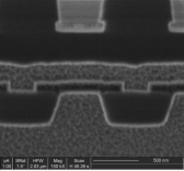
The electron microscope consists of several components, including the specimen stage, the electron beam, and the detector. The specimen stage is where the samples or objects to be examined are placed. The electron beam is used to excite the electrons in the specimen, and the detector measures the electrons that are emitted or scattered from the specimen.
The specimen stage can be made of various materials, such as glass, plastic, or metal. It is important to choose a material that is compatible with the type of specimen being examined. The electron beam can also be directed at the specimen using various techniques, such as by focusing it on a single point or by using a beam of electrons that is deflected at a specific angle.
The detector is used to measure the electrons that are emitted or scattered from the specimen. There are several types of detectors that can be used, including the camera, the analyzer, and the image intensifier. The camera detects the electrons and converts them into an image that can be displayed on a screen. The analyzer is used to control the beam of electrons and to focus it on the specimen. The image intensifier is used to increase the contrast and resolution of the image.
In addition to the components, the electron microscope also has various options and techniques that can be used to improve the accuracy and quality of the image. For example, the microscope can be equipped with an optional stage that allows for the examination of thin films or biological samples. It can also be equipped with a high-resolution transmission electron microscope, which allows for even higher magnifications and resolutions.
In conclusion, electron microscopy is an important tool in modern science, with applications in fields such as materials science, biology, and medicine. It allows researchers to study the structure and behavior of molecules and cells at the atomic and subatomic level. The microscope consists of several components, including the specimen stage, the electron beam, and the detector, and various techniques and options can be used to improve the accuracy and quality of the image.

上一篇
分析电镜英文